
Tim Vernimmen in Nautilus:
 You don’t often see the word beautiful in scientific articles. Yet it’s easy to see why cell biologists Niccolò Banterle and Pierre Gönczy used the word when describing a crucial cell structure called the centriole in a recent review. The scientists, of the Swiss Federal Institute of Technology Lausanne, are helping to provide a more precise view of how our cells construct this microscopic marvel.
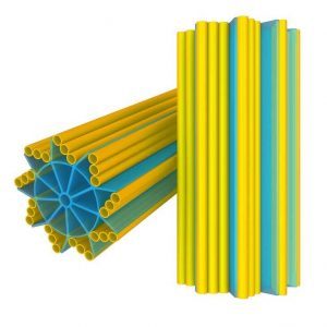
Seen in the image in cross section, the centriole is a cylindrical cell organelle built of nine elegant groupings of three hollow tubes known as microtubules. Each centriole is about one-300th the width of a thickish human hair (about 250 nanometers) in diameter and as much as twice as long. It sits in an area of the cell that looks distinctly fuzzy under the microscope; that fuzzy spot plus the centriole are together known as the centrosome. Each human body contains trillions of them. Centrosome disorders have been implicated in a variety of birth defects, such as certain forms of dwarfism and microcephaly, and may have a role in cancer. Centrioles have existed for over two billion years and are found in a wide variety of life forms that may use them in different ways. In single-celled organisms such as Euglena or Trypanosoma brucei (the parasite that causes sleeping sickness), centrioles form the core of a flagellum, a whipping appendage that enables cells (including human sperm cells) to propel themselves. That’s probably the job that centrioles first evolved to do, says Gönczy. Over time, though, the same structure was adapted for another function: fine-tuning the orderly division of cells. Every one of us starts out as a single fertilized egg cell, which then divides into two daughter cells that each produce two daughters of their own, and so on. Getting enough divisions done in the right way and in adequate time is crucial for correctly structuring developing tissues.
More here.